Wednesday, February 29, 2012

sperm mediated transfer 2012


Eliane's thesis is available on line.

Manipulation of Sperm for Efficient Production of Transgenic Calves and Chicks.
Eliane Harel- Markowitz


Abstract
The current method of micromanipulation used in domestic animals results in less than 1% transgenic animals, which makes it extremely difficult to produce transgenic calves and is completely unfeasible for producing transgenic chicks. The purpose of this work was to find a more efficient method for producing transgenic calves and chicks using a combination of two techniques, lipofection and restriction enzyme mediated insertion (REMI). Previously investigators were unable to produce transgenic chicks using lipofection alone, while injection of isolated sperm nuclei, which had been incubated with restriction enzyme, into oocytes had only been demonstrated in frogs. In this study, we demonstrated for the first time, that lipofection of both DNA and restriction enzyme could be used to successfully integrate DNA into sperm genome DNA and then used for routine AI to produce transgenic calves and chicks. First it was demonstrated using needle pricking and southern blot analysis of genomic DNA that the restriction enzyme opens up “hot  spots in the sperm genomic DNA, producing sticky ends into which foreign DNA  could be inserted and integrated into the sperm genomic DNA rather than just free DNA floating in the fertilized egg. The “transgenic sperm” thus made were used in IVF and AI to produce embryos expressing a foreign DNA, GFP (green fluorescent protein). Using Not I and linearized pGFP lipofected to sperm for AI resulted with two calves which expressed the exogenous DNA in their lymphocytes as determined by (a) PCR and RT-PCR; (b) specific emission of green fluorescence by the GFP protein; (c) homology analysis between GFP DNA and PCR product DNA sequences and (d) Southern blot analysis.  Similarly in the chicken, linearized plasmid GFP sequences with the corresponding restriction enzyme (REMI) were lipofected into the sperm. The transfected sperm were then used for AI in hens and 90% (17/19) of the resultant chicks expressed the exogenous DNA in their lymphocytes as determined by  (a) PCR and RT-PCR; (b) specific emission of green fluorescence by the GFP and (c)  Southern blot analysis. A complete homology was found between the Jellyfish GFP DNA and a 313 bp PCR product of DNA from chick blood cells. The procedure was then tested with an additional construct, hFSH. The construct of hFSH consisted of both subunits, alpha and beta and the PCR product used primers for both alpha and beta subunit resulted with a PCR product of 584 bp which was unique to transgenic chickens. The procedure was then used to lipofect a construct of hFSH (Human Follicle Stimulating Hormone) into chicken sperm and used for AI. The resultant offspring were transgenic in at least three generations as determined by (1) measurement of hFSH protein in chicken blood using enzyme immunoassay (EIA) and RIA;  (2) RT-PCR and PCR; and (3) copy number.  We conclude that (1) lipofection of both DNA and restriction enzyme into sperm  (bovine and chicken) induces the integration of the DNA into the sperm genomic DNA; (2) lipofected sperm can be used in AI to produce a high percentage of transgenic calves and chicks; (3) The integrated gene is expressed in first, second and third generation; and (4) the method is not limited to specific genes. The technique of lipofection of DNA combined with REMI is therefore an efficient and stable method for producing transgenic domestic animals.  Efficient production of transgenic domestic animals could have major impact on gene therapy, improving livestock breeds and the production of valuable pharmaceuticals, e.g. hFSH, which could be extracted from eggs and milk.


The e-book Sperm-mediated Gene Transfer: Concepts and Controversies. (ed. K. Smith) is  online.


Sperm-Mediated Gene Transfer: Concepts and Controversies
DOI: 10.2174/97816080523701120101
eISBN: 978-1-60805-237-0, 2012
Editor: Kevin R. Smith  Abertay University

Manipulation of Sperm for Efficient Production of Transgenic Calves and Chicks Pp.92-102
Mordechai Shemesh, Laurence Shore, Yehuda Stram, Eliane Harel-Markowitz and Michael Gurevich

Introduction

Sperm-mediated gene transfer (SMGT) represents a novel set of technologies for animal (or in the future, human) genetic modification using the sperm as a vector, as opposed to more traditional established routes such as fertilized eggs or embryonic stem cells.

Studies of sperm-mediated gene transfer (SMGT) indicate that sperm cells possess the ability to be utilized as carriers of exogenous genetic sequences, offering the potential of a novel cost-effective route for germline genetic modification. The fate of transgenes borne by sperm cells has been inconsistent, and analysis of offspring from SMGT experiments has shown a mixed picture in terms of genomic integration of the transgene, suggesting an episomal mode of inheritance. Various distinct steps in transgene uptake by the sperm cell have been described or proposed, including a model based upon endogenous reverse transcriptase activity. Although mature sperm cells are naturally protected against uptake of foreign nucleic acid molecules, certain environmental conditions, for example at key times within the reproductive tract, may reduce this protection, suggesting that SMGT may occasionally take place in nature. If correct, this carries profound implications for evolution and human genetic health. This e-book brings together theoretical and empirical reviews from experts in SMGT, providing comprehensive coverage of the major trends, developments and controversies in this novel field. This e-book is intended as a reference for professional researchers in the field of animal genetic modification (transgenesis) as well as teachers, scientists and physicians interested in medical genetics in general and gene therapy in particular.


Shemesh, M, Shore L., Stram Y., Harel-Markowitz, E. and Gurevich M. (2012). Manipulation of sperm for efficient production of transgenic calves and chicks. In: Sperm-mediated Gene Transfer: Concepts and Controversies. (ed. K. Smith) pp. 130-145. Bentham E-book


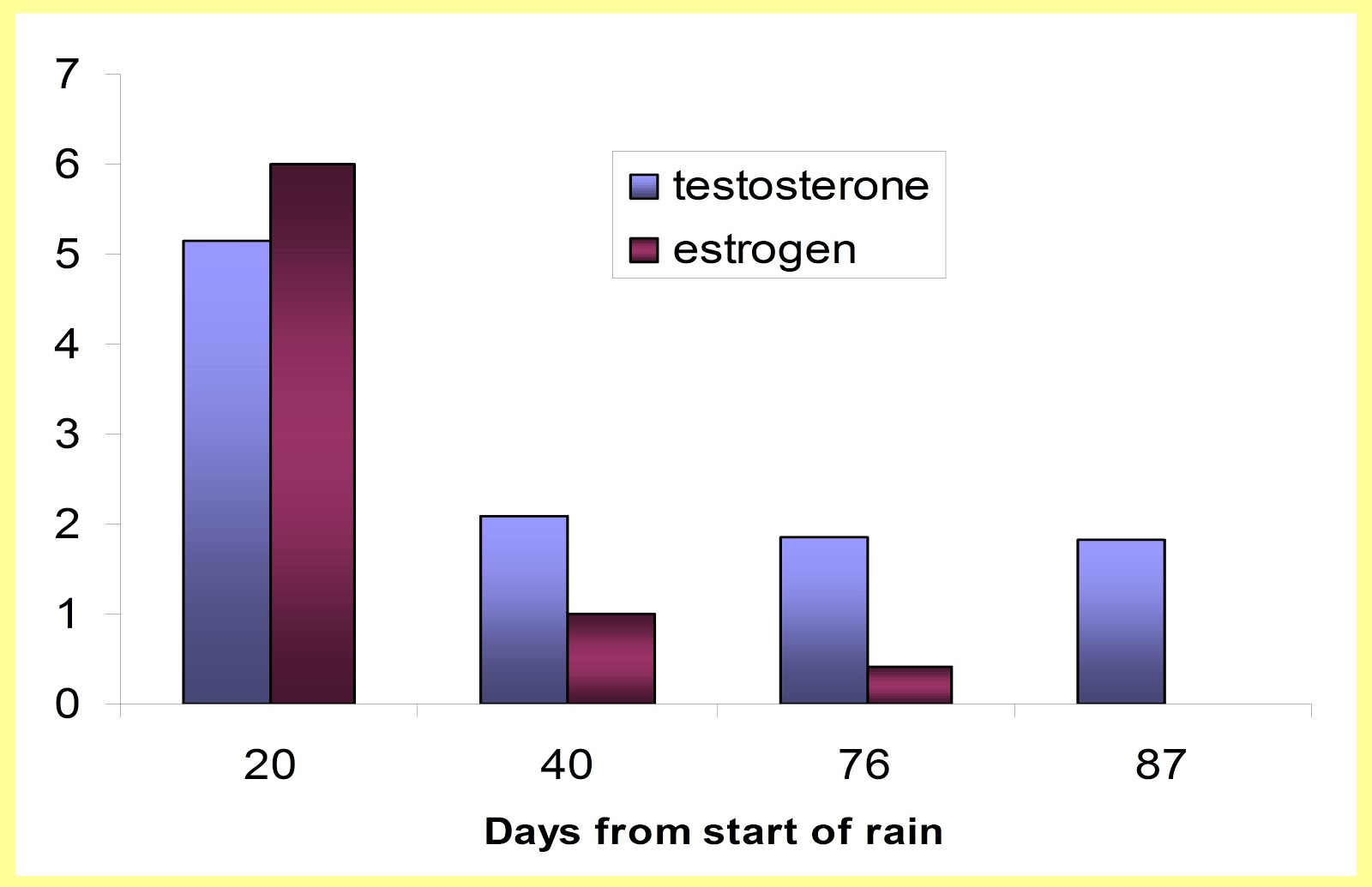
The current method of micromanipulation used for domestic animals results in less than 1% transgenic animals. This makes it extremely difficult to produce transgenic cows and is not feasible for producing transgenic chickens. The purpose of this work was to find a more efficient method for producing transgenic calves and chicks using a combination of two techniques, lipofection and restriction enzyme mediated insertion (REMI). Previously investigators were unable to produce transgenic chickens using lipofection alone. On the other hand, injection of isolated sperm nucleus incubated with restriction enzyme into oocytes has only been shown to be effective in frogs. In this study, we demonstrated for the first time, that lipofection of both DNA and restriction enzyme could be used to successfully integrate DNA into the sperm genome DNA and then used for routine AI to produce transgenic calves and chicks. First it was demonstrated using needle pricking and southern blot analysis of genomic DNA that the restriction enzyme opens up “hot” spots in the sperm genomic DNA. This produces sticky ends by which foreign DNA can be inserted and integrated into the sperm genomic DNA. The “transgenic sperm” thus made were used in IVF and AI to produce embryos expressing a foreign DNA, EGFP (enhanced green fluorescent protein). Using Not I and linearized pEGFP lipofected to sperm for AI resulted with two calves which expressed the exogenous DNA in their lymphocytes as determined by (a) PCR and RT-PCR; (b) specific emission of green fluorescence by the EGFP protein; (c) homology analysis between EGFP DNA and PCR product DNA sequences and (d) Southern blot analysis. Similarly in the chicken, linearized plasmid EGFP sequences with the corresponding restriction enzyme (REMI) were lipofected into the sperm. The transfected sperm were then used for AI in hens and 90% (17/19) of the resultant chicks expressed the exogenous DNA in their lymphocytes as determined by: (a) PCR and RT-PCR; (b) specific emission of green fluorescence by the EGFP; and (c) Southern blot analysis. A complete homology was found between the Jellyfish EGFP DNA and a 313 bp PCR product of DNA from chick blood cells. The procedure was then tested with an additional construct, hFSH. The construct of hFSH consisted of both subunits, α and β and the PCR product used primers for both α and β subunit resulted with a PCR product of 584 bp which was unique to transgenic chickens. The procedure was then used to lipofect a construct of hFSH (Human Follicular Stimulating Hormone) into chicken sperm and used for AI. The resultant offspring were transgenic for at least three generations as determined by: (1) measurement of hFSH protein in chicken blood using enzyme immunoassay and RIA; (2) RT-PCR and PCR; and (3) copy number.
We conclude: (1) that lipofection of both DNA and restriction enzyme into sperm (bovine and chicken) induces the integration of the DNA into the sperm genomic DNA; (2) lipofected sperm can be used in AI to produce a high percentage of transgenic calves and chicks; (3) The integrated gene is expressed in the first, second and third generation; and (4) the method is not limited to specific genes. The technique of lipofection of DNA combined with REMI is therefore an efficient and stable method of producing transgenic domestic animals. Efficient production of transgenic domestic animals could have major impact on gene therapy, improving livestock breeds and the production of valuable pharmaceuticals, e.g. hFSH, which could be extracted from eggs and milk.


Real time RT-PCR for hFSH from second generation transgenic chicken tissues using 1:10 serial dilutions of 0.2 µg/tube of extracted RNA.
The sample amplification curves begin rising between the 35th and 39th cycle of PCR, while negative control (not shown) remained horizontal. The differences in the cycle no. of the curves represent differences in the gene expression in different tissues


Wednesday, February 15, 2012

Environmental estrogens 2012


The Israel Journal of Veterinary Medicine has issued a special issue in honor of Prof. Natan  Ayalon
here


This is the abstract of our contribution.


Effects of Environmental Estrogens on Reproductive Parameters in Domestic Animals
Shemesh, M. and Shore, L.S. Department of Hormone Research, Kimron Veterinary 
Institute, Bet Dagan, POB 12, 50250 Israel.


ABSTRACT

Environmental estrogens are natural products of plant (phytoestrogens) or animal origin (steroidal  estrogens) that have estrogenic properties. They are a major group in a category known as endocrine disruptors. In the field of animal husbandry, the effects of these environmental estrogens are well documented. This paper discusses  the effects of plant estrogens and environmental steroid hormones, such as estrogen and testosterone, as seen in animals in Israel. The areas considered are reproductive disorders, premature udder development, prolapsed oviduct, scrotal atrophy and skewed sex ratios.
Keywords: phytoestrogens, zearalenone, estradiol 17-β, testosterone, testes, oviductal prolapse, scrotum, sex ratio
Israel Journal of Veterinary Medicine  Vol. 67 (1):6-10 March 2012


Effect of estrogen implants on scrotal circumference and tonus in bull calves. Thirteen calves were implanted at 3 mo and their testicles measured at 6 months. Fourteen other calves similarly fed and housed served as controls.  The control circumference (cm) was significantly larger than the treated (P<0.01) and the tonus was significantly lower in treated bulls (P<0.02; 10=very poor; 20=poor; 30=good).


There is scant literature on the effects of exogenous estrogen on bull fertility.  Here is the graph from a short note  that I found.
Infertilité mâle bovine et teneur en coumestrol de la Luzerne.
Infertility in bulls and the concentration of coumestrol in alfalfa.
J.Le Bars, Nurard J.S.  Pharmacologie et Toxicolgie Vétérinaires, INRA Publ. Paris 1982 
Le Colloques de l'I.N.R.A. 8:157-158


 Belgian Blue or Belgian White Blue Bull. Established in the early 20th century, this heavily- (double) muscled, mottled Belgian breed, originally intended for beef and dairy use, is now mainly a beef breed, with bulls usually crossed as terminal sires to produce fast developing veal calves.
(cfgphoto.com copyright, used with permission)

For some reason, the picture of the Belgian Blue is getting a lot of hits.  Here are some more of  7 month old Belgian Blue calves selected for an insemination program taken in 1986 in Belgium (courtesy of Dr. S. Marcus)
Belgium blue calf 01


Belgium blue calf 02


Established in the early 21th century, this heavily-double muscled, mottled breed, originally intended for a scientific experiment. is now a  sporadic superhero.

Cleaning out the drawers and found this little note.

 Pregnancy in a hermaphroditic cow.  High estrogen content of the hermaphroditic testes .
Perl, Smuel; Marcus, Smuel; Shore, Laurence S.; Brenner, Gideon;  Shemesh, Mordechai
A pregnant heifer was examined after it was observed to have masses in the pelvic region.  At slaughter, it was found to have a gravid uterus with a 3 month old fetus.  Testes were observed in the mesovarium .  One of the testes was well formed with an epididymis and seminal vesicular structure. Histological examination of the testis showed Sertoli cells, Leydig cells, germ cells and seminal  vesicles.  The second testis was small and contain only medullary cell mass.  Steroid analysis of the formed testes indicated high levels of estrogen and normal levels of testosterone compared to normal testes [Table 1].
There has been a least one previous report of a pregnant hermaphroditic cow. [Trächtigkeit bein einem Zwitter des Rindes (Hermaphroditaismus verus alternans). Fürst.   Deutsche Tierärztliche Wochenschrift  121 15. April.  59:121, 1952] .   Estrogen has long been known to be present in bull seminal fluid [Payne A, Kelch R, Musich S and Halpern M. Oestrogen content of semen and the effect of exogenous oestradiol-17β on the oestrogen and androgen concentration in semen and blood plasma of bulls. Journal of Reproduction and Fertility  50:17-21 (1977)] and aromatase can be present in all of the various cells of the bovine testes. [Carreau, S.; Hess R.A.  Oestrogens and spermatogenesis Phil. Trans. R. Soc. B 2010 365, 1517-1535.]


Tissue source
Testosterone
Estrogen

ng/g  fresh wt
ng/g  fresh wt
Hermaphrodite


Interstitial
35.0
2.0
Leydig
8.2
0.3
Epididymal
33.1
0.4


Normal Calf (1 yr)


Interstitial (left)
42.0
<0.1
Interstitial (right)
60.5
<0.1
Epididymal
2.8
<0.1
Epididymal
4.0
<0.1


Estrogen binding receptor 

For many years  the Hormone Research Laboratory made their own cytosolic estrogen receptor.  It was used for the determination of phytoestrogens like coumestrol in alfalfa and zearalenone in corn products.  However, it became  too difficult to  obtain 6 dy old pregnant rabbits so we abandoned the assay.  Efforts to find a replacement have not been successful to date. The most promising system is XDS's Lumi-cell ER based on engineered cells linked to luminiferase.


Seems to work nicely for zearalenone, coumestrol, genistein and diadzein in the expected order.
The upper graph also shows alpha estradiol which usually isn't tested for.  Alpha estradiol is the principal estrogen found in cow blood but is not considered an active estrogen. 

I guess I should mention invitrogen which makes LanthaScreen™ TR-FRET Estrogen Receptor alpha and beta assays.  On paper this sounds impressive.  An assay using recombinant receptor linked to the  rare earth element terbium.  When the receptor is engaged, the terbium shifts its spectrum which can be detected with a spectrophotometer.   They do not offer any training or someplace where you can go to see it in action.  After I ordered it and went over the specs, I saw that it could not possibly work on the spectrophotometer I had even though it stated on their web site that it would work.  After a short email to the company, I was informed that indeed it would not work.  If somebody out there wants the kit, they are welcome to it.  Google states not to put your email on a web site because of spammers so just write me on Facebook or something.  Laurence Shore


Media


Originally published August 18, 2012 at 4:41 PM | Page modified August 18, 2012 at 5:20 PM

Israel sperm banks find quality is plummeting
The quality of the product being offered to Israeli sperm banks is falling at an alarming rate, and no one is sure why.

By Edmund Sanders
Los Angeles Times


JERUSALEM — The founder of the Tel Aviv-based specialty firm raves about his product with the same gusto distillers reserve for their top-notch scotch. He's particularly proud of his "premium" line. Sure, it costs a bit more, but it's targeted at a more discriminating client.
Dr. Jacob Ronen is in the sperm business. Among other things, as head of Cryobank Israel, the country's largest private sperm bank, he guarantees that his stable of superior donors includes only tall, twenty-something ex-soldiers whose sperm has passed rigorous genetic testing.
But finding such super sperm isn't as easy as it used to be. Only 1 in 100 donors makes the cut. A decade ago, it was 1 in 10.
It's not just first-rate sperm that's in short supply. All of Israel's half a dozen or so sperm banks are scrambling to keep their liquid-nitrogen freezers stocked.
Simply put, the quality of Israeli sperm is falling at an alarming rate, and no one's sure why.
Fertility is a major issue in Israel, where memories of the Holocaust genocide are fresh, and having children is an entrenched part of Judaism. There's also a political aspect because birthrates among Arabs in Israel have at times been as much as double those of Jews, triggering a population race that some believe could one day affect who controls the land.
So the drop in the quality of sperm is raising some red flags, even though the cause remains a mystery. Theories range from the mundane (carrying cellphones in front pockets) to the far-fetched (depleted uranium from exploded munitions). Some Israeli scientists are looking at naturally occurring hormones, particularly estrogen, in Israel's water and milk. They suggest it's a mark of the country's aggressive dairy-farming methods.

In the microscope

The director of the Hadassah Sperm Bank, Ruth Har-Nir, hunches over a microscope to view a freshly donated specimen and begins to methodically count each squiggly swimmer magnified on the slide.
She is checking the quality of a prospective donor, a young graduate student hoping to earn extra cash. Though sophisticated lab machines could be used to analyze potency, Har-Nir says the old-fashioned method works best.
After a quick scan, she sits up and shakes her head. The number of spermatozoa darting around each tiny grid on the slide is two to four, well below the minimum six required, and nowhere near the 10 to 20 per grid that indicates the concentration the bank likes to see.
Also, rather than surging forward, some of the little guys flit left and right or just stall out, suggesting a weak motility.
"Under no circumstances can we accept sperm of this quality," she says. In the previous three weeks, her bank tested six candidates and rejected all. "This is the trend," she adds.
When Har-Nir helped start the sperm bank in 1991, she says, it turned away about one-third of applicants because of low quality. Using the same standard today, it would reject more than 80 percent. Though the bank relaxed its criteria, it still vetoes about two-thirds.
Long-term change
Har-Nir noticed the problem a decade ago when she began rejecting more and more sperm from otherwise healthy young men. She shared her observations with local fertility doctors, and their research has confirmed her suspicion.
In the past 10 to 15 years, the concentration of sperm samples collected by the bank dropped 37 percent from 106 million cells per milliliter to 67 million, said Dr. Ronit Haimov-Kochman, a leading Israeli infertility researcher at the Hadassah Hebrew University Medical Center.
Though declining sperm quality is an international phenomenon, the change in Israel is occurring at nearly twice the pace as other developed countries, Haimov-Kochman said. If current trends continue, she said, by 2030 the concentration of sperm from Israeli donors will drop below 20 million cells per milliliter, which many international health experts define as abnormal.
There's no evidence declining sperm quality is resulting in fewer babies. The birthrate of Israel's Jewish population has risen in recent decades, thanks largely to an increase in the number of ultraorthodox Jews, who tend to have large families.
Haimov-Kochman estimated infertility rates in Israel have risen from 10 to 15 percent in the past 15 years, but said that's in line with international trends. But she said male infertility — once believed to be the cause about half the time, just as in the U.S. — is now suspected in 70 percent of the Israeli cases.
Most worrisome, she added, is that research has focused on sperm-bank donors, mostly students who are younger and healthier than the general population.
"If this is happening to the guys on our A-team, we might only be seeing the tip of the iceberg," she said.

Banks reaching out

Sperm banks are struggling to cope. Rather than rely on walk-ins as they once did, they use marketing campaigns, posters in college sports centers and Facebook pages to attract virile candidates. The going rate for a donation has doubled in the past 10 years to about $270.
"The decline (in sperm quality) has been dramatic," Cryobank's Ronen said. "It's a shame. We see these macho, beautiful guys come to give donations, but then we're embarrassed to have to tell them that their sperm quality is so low they may actually end up coming back as a client."
He's capitalized on that, though, by offering to freeze sperm of young men with borderline quality who want to set aside a reserve in case their potency declines with age.
Har-Nir said her bank sometimes refers men with the most serious deficiencies for counseling or medical advice. But she emphasizes that rejection by the sperm bank doesn't necessarily mean they won't be able to have children naturally. It just means their sperm isn't, well, commercial quality.

Too much estrogen?

Even with the drop in sperm quality being well-documented, the cause remains unclear and the theories controversial. Some scientists fear Israelis are being overexposed to female hormones.
"People in Israel are getting quite a load of estrogen," said Laurence Shore, a retired hormone and toxicology researcher at the Kimron Veterinary Institute near Tel Aviv. "I don't think it's a good idea to expose children to such high levels of estrogen."
He said no studies have determined estrogen levels in Israel are harming humans, adding that exposure may be too low for that. But he said it might be a factor in the sperm decline.
His research has found Israeli milk and associated products such as butter and cheese can contain 10 times as much estrogen as products from other countries because of Israel's aggressive milk-production practices.
Israel is a world leader in producing milk, pumping twice as much from its cows as other parts of the world, he said. That's partly because cows are milked up to their eighth month of pregnancy, when natural estrogen levels in the milk soar, according to Shore. In nature, he said, cows usually stop giving milk to their own young when they are three months' pregnant with a new calf.
Even though many other nations have adopted similar milking practices, Shore said, Israel is one of the first and most aggressive, so it could be seeing the effect sooner.
Haimov-Kochman is looking into water quality. As a tiny nation with a shortage of water, Israel reclaims much of its used water and sewage, which is processed, used in agriculture and may find its way back into groundwater.
The water, she said, has been found to contain traces of ethinyl estradiol, a synthetic estrogen used in birth-control pills, which gets into the water through the urine of women taking the pills.
"You can't clean this from the water," she said.
Haimov-Kochman is also studying the effect of phthalates, chemicals used in plastic products that are suspected of affecting male reproductive development.
"But I can't prove any of this," she said.
Industry and government scientists dismiss fear about Israel's water and milk as unfounded, saying levels are too small to affect humans.
"Only a tiny part of the total estrogen produced by the cows ends up in the milk," said Dr. Stefan Soback, director of the Ministry of Agriculture's National Residue Control Laboratory. "It is not sufficient to determine estrogen content in milk in order to claim physiological effects to somebody that consumes it."

Batsheva Sobelman of the Los Angeles Times contributed to this report.

Some comments:


"Although estrogenic hormones in milk from nonpregnant cows are in the pg/ml range, milk from pregnant cows can contain 500 ng estradiol/l, 1 µg estrone/l (mostly as conjugated sulfate), and 10 µg progesterone/l [43] (and approximately half of dairy herd is in late pregnancy during milking). Human breast milk for infants contains little estrogen or progesterone since nursing humans are generally not pregnant. Since young children consume about 300–700 ml/day of bovine milk, they may ingest 40 to 100 ng/day of estrogen (estradiol, estrone, and estrone sulfate), and whether this can be considered a safe level is a matter of debate [45]. "

http://old.iupac.org/publications/pac/2003/pdf/7511x1859.pdf

Shore L.S. and Shemesh M. (2003). Naturally produced steroid hormones and their release into the environment. Pure and Applied Chemistry 75:1859–1871


In my opinion, 0.1 microgram per day is not a negligable dose since the daily maintanance level for post-menapausal women is 25 microgram/day. Mice will respond to 50 ng/day of estradiol (A.Kaye, personal observation). I was at the cow meeting a few years ago and some enterprizing young man wanted to extend miliking to 7 and half months. I objected strongly at the time and I trust it was never put into practice. 


Health Effects of Cows’ Milk
http://www.eps1.comlink.ne.jp/~mayus/eng/
Commercial cows’ milk has estrogenic activity as revealed
by the hypertrophic effects on the uteri of young
ovariectomized rats and immature rats
Adapted from:
Ganmaa D, Tezuka H, Enkhmaa D, Hoshi K, Sato A.
Commercial cows’ milk has uterotrophic activity on the uteri of young
ovariectomized rats and immature rats.
International Journal of Cancer 2006;118:2363-65.
Abstract
Cows’ milk has considerable amounts of estrogens, mainly in the form of estrone sulfate. To determine whether the commercial milk has any biologically significant hormonal effects, two series of uterotrophic tests were performed, one with young ovariectomized rats and the other with sexually immature rats. Thirty-six rats were used for each test. They were divided into 3 groups of 12 animals each, and were kept for 7 days on powdered chow with one of three drinking solutions: low-fat milk (experimental), artificial milk (negative control), or artificial milk containing estrone sulfate at 100 ng/ml (positive control). At autopsy, both the wet and blotted uterine weights were measured. The cell heights of uterine epithelia in ovariectomized rats were also determined. In each test, the weights of the uteri in the Low-Fat Milk group were significantly greater than those of the respective weights in the Artificial Milk group (p<0.01). Furthermore, in ovariectomized rats, the uterine epithelial-cell height in the Low-Fat Milk group was significantly greater than that observed in the Artificial Milk group (p<0.01). The uterotrophic effect of 100 ng/ml Estrone Sulfate solution was greater than that of Low-Fat Milk in immature rats (p<0.01), whereas the effect of the solution was almost comparable to that of Low-Fat Milk (p>0.05). In conclusion, commercially available low-fat milk has uterotrophic effects in both young ovariectomized rats and sexually immature rats.


 The standard in US and England is that cows are milked till 220 days or 60 dry days before calving.  Cows are pregnant the same amount of time as humans (283 days and have the same development rate, i.e. differentiation of the sexual organs occurs between 45-90 days of gestation.)


Tuesday, February 14, 2012

environment hormones in hula valley

This was a chapter for a book on the Hula that was never published.

Source and transport of natural and synthetic steroid hormones in Hula Valley and their  effects on fish in aquiculture.
L. Shore*, Ph.D, Kimron Veterinary Institute, Bet Dagan, POB 12, Israel
 Key words:  testosterone, ethinylestradiol, estrogen, Cyprinus carpio 
Abstract
Natural and synthetic steroid hormones are constantly being excreted into the environment.  In the Hula Valley, five steroidal compounds were identified in the surface waters – testosterone, estrogen (estradiol-17b and estrone), androstenedione, ethinyl estradiol and estriol.  The sources of the hormones were shown to be cattle pasture and treated sewage effluent and the sources could be identified based on hormone profile.  However, of the five steroids tested, only testosterone could permeate the soil to reach the groundwater.  Fish with male gonadal atrophy but normal female gonads were found in a reservoir used for aquaculture. The gonadal atrophy was associated with the presence of hormones, specifically the synthetic hormone, ethinyl estradiol, which is used in contraceptive pills.    When in the subsequent year, the same fish were grown in the absence of the hormone, the males were found to have normal gonads.  
Introduction
In the past decade, many concerns have been expressed on compounds that mimic hormones and can disrupt reproduction in animals including humans (see Lintelmann et al., 2003 for review).  Of particular concern are synthetic hormones that are specifically design to act on biological systems.  All of these compounds act against a background of naturally produced hormones such as testosterone and estradiol that are constantly release into the environment (see Shore and Shemesh, 2003 for review).  Since our initial observation of the presence of significant amounts of testosterone and estrogen in Lake Kinneret in 1993 (Shore et al. 1993), we have surveyed the Jordan Valley including the Upper Jordan River Catchment and the Southern Jordan River for the presence of steroidal hormones (Bar-El Cohen et al., 2005; Shore et al.. 2004).   The present paper will described the results of these surveys in the Hula Valley as to the determination of sources, the pathways of transport and the effects on fish. 
The primary steroid hormones excreted into the environment are estrone, estradiol-17β, testosterone and androstenedione (Fig. 1). The natural steroids of major concern are estrone and estradiol since they exert their physiological effects at lower concentrations than other steroids and can be found in the environment in concentrations above their Lowest Observed Effect Level (LOEL) for fish (increased vitellogenin) and plants (increased growth) (10 ng/l) (Christiansen, 2002; Shore et al., 1992,1995b). In rivers and soil, estradiol is converted abiotically to estrone, so for environmental studies estradiol and estrone can be considered together as “estrogens” (Colucci et al., 2001; Jürgens et al., 2002). Effluent from human sources can also contain estriol, a weak estrogen excreted in the urine of pregnant women, and synthetic estrogens such as mestranol and ethinylestradiol (Fig. 1) (Wenzel et al., 1998). These synthetic compounds are of particular concern as they have LOEL’s in the order of 1 ng/l (Christiansen et al., 2003).
To study the occurrence, origin and effects of the natural and synthetic steroids, a general survey of the Hula Valley catchment area was performed as well as more in depth studies, at two sites to (1) determine nature of export of hormones from cattle pasture and (2) determine the effect of the hormones on fish raised in aquaculture.
Materials and methods:

Extractions and assays

Manures: Extraction and determination of testosterone, and estrogen in cattle manure were done by established procedures (Shore and Shemesh, 1993; Shore et al., 1993). Androstenedione was determined using a commercial ELISA (DRG GmbH, Marburg, Germany). 
Water: All water samples were collected into acid-washed polyethylene 1 L bottles, and immediately put in a cooler before transporting to the laboratory for further processing.  Extraction and determination of testosterone, estrogen, estriol and ethinylestradiol were done as previously described (Shore et al, 2004).
Soil:  Five gram samples were extracted twice with 15 ml of ethyl acetate, the supernatants combined, evaporated and redisolved in 0.5 ml of methanol.   100 µl aliquots were evaporated to dryness and redisolved in the testing media for analysis.
Statistics: All data are in means ± SD. Student t test used for comparing two groups.  


Figure 1.  Structures of natural and synthetic environmental steroids

Site description
The Hula Valley catchment area is divided by the Jordan River into a Western and Eastern sub-catchment areas.  The Eastern portion (Golan Heights, eastern part of the Hula valley) is characterized by the confluence of the Dan, Hermon and Sneir tributaries at the Joseph Bridge to form the Jordan River, which receives the runoff from the Golan Heights until it leaves the Hula Valley at a second confluence, the Huri Bridge.   At the Joseph Bridge, about 10% of the Jordan River is diverted to the Western Canal which receives runoff from the western or Naphtali Heights.  The Western Canal rejoins the Jordan shortly before its exit from the Hula Valley. Precipitation in the Hula Valley catchment area occurs only in the rainy season from Oct. to May. The area consists of small farms, cattle pasture, fishponds with some urban development.  The water table is shallow being less than 2 meters from the surface. The fifteen sites sampled after each rain event (11 to 12 times a hydrological year) on the eastern side of the catchment are shown in Figure 2.  The sites samples on the western side were in two separate subcatchments (points P, KD on  Fig. 3)  which were sampled on two occasions (Jan. 24, Feb 2, 2005) following rain events. For the aquiculture observations, a reservoir containing 400 tons of three species of fish (grey mullet, carp and tilapia) was sampled to determine the effect on aquaculture of exposure to sewage effluent (site 404/2, fig. 3).  The density of the fish was approximately 1 fish/l.  The reservoir held 5.6 mcu3 over area of 100 acres and a depth of 15 m. The reservoir received water from three sources: sewage effluent from an urban area of about 8000, excess water flow from Lake Hula  and Jordan River water from the Western canal. The limonological properties of the reservoir have been described in detail (Gafny et al., 1999). Samples were taken on 16.02.05 before fish were introduced and 04.05.05, 04.08.05 and 19.11.05 representing 1, 5 and 8 months after the introduction. Samples were taken at depths of 1, 3 and 6 m except on the final sampling before harvesting when the depth was only 0.8 m. Samples were taken from a control reservoir without fish on the same dates.  In Nov. 2005, the remaining males were transferred to another pond containing the same water without any sewage contaminant. Gonadal somatic index (GSI) for the carp was evaluated according to Degani et al. (1996, 1998).
For the cattle pasture study, a fenced field in which cattle were not present most of the year with a pool receiving runoff from the field was studied after the introduction of the cattle for three months. The grazing intensity was 0.63 cows/ha. For edge of field drainage, one liter samples in polyethylene bottles were obtained from the receiving pool after each of three rain events. Samples were acidified and transported to the laboratory.  Single samples of soil were taken after 1 mo., 2 mo and 3 mo. after introduction of cattle at a depth of 10, 20 and 30 cm.

Figure 2.  Sites in the Hula Valley draining the Eastern side of the catchment sampled in the hydrological years 2001/2 and 2002/3. The sites are listed in Table 1 found below.


Figure 3. Two subcatchments (Di, Kd) draining cattle pasture in the western portion of Hula Valley catchment studied in hydrological year 2003/2004.  The defined cattle pasture studied for soil and manure samples was located at N2 (Site #1). The reservoir used for aquaculture was at site 404/2.


Results
Eastern part of the Hula Valley
The transport of testosterone, estrogen, ethinylestradiol and estriol was measured at fifteen sites in the eastern side of Hula Valley catchment area after major rain events (10 -12 per hydrological year).  It was found in the rain season of 2001/2002, that following a rain sequence of 131 mm/wk, there was an initial large increase in the concentration of testosterone (maximum 6 ng/l) accompanied by high estrogen (maximum 6 ng/l), which then gradually declined to non-detectable levels (<0.3 ng/l) over a period of three months. These peaks originated from runoff from cattle pasture (Fig. 5). Later peaks consisted only of testosterone that was moderately associated with sulfate (r2=0.53, P<0.05) and somewhat associated with total phosphorus (r2=0.49, P<0.1) indicating that the origin was leaching from the sulfate rich peat soil typical of the Hula Valley (Fig. 6). Testosterone in this catchment area was therefore due initially to surface runoff from cattle pasture and then as discharge from the soil.  The testosterone pulses after each rain event were more pronounced at the Joseph Bridge than at the Huri Bridge (Fig. 7).  Ethinylestradiol was consistently above 1 ng/l at three sites, two of which were known to receive sewage effluent and one had a high ammonia (>1.2 mg/l) and bacteria count (>50,000 fecal coli/100 ml), probably originating from a leaky sewage holding pond. There was no correlation (r<0.2) with the amount of testosterone, ethinylestradiol, estrogen or estradiol present in the samples with each other. However in the few sites (5/28) in which estriol was above 1 ng/l, ethinylestradiol was also above 1 ng/l (15/28).  A more detailed report on the results for 2001/2002 and 2002/2003 hydrological years can be found in Shore et al., 2004.


                     Ng steroid/l        


Figure 4.  Hormonal profile of runoff from cattle pasture.  


Figure 5.  Testosterone and sulfate concentrations in the runoff in the Hula Valley after rain events in the 2001/2002 hydrological year.
Table 1. Representative testosterone and ethinylestradiol concentrations at 14 sites on the Eastern side of the Hula Valley.
Site no.
Site name
Testosterone
ng/l
Ethinylestradiol
ng/l
Comments


1
Dan Tributary
1.1
1.3


2
Hermon Tributary
0.9
1.1
Some raw sewage
3
Sneir tributary
1.4
0.7


4
Hyun Creek
1.1
0.9

5
Joseph bridge
2.0
0.5
Confluence of sites 1,2,3,4
6
Kalil Canal
1.3
0.8


7
Yardonin Creek
2.1
1.2


8
Gonen Canal
2.2
1.6
High ammonia and fecal coli
9
Bitachon Canal
1.4
1.3


10
Lehavot Bridge
1.2
0.7
River below sites 6,7
11
Green Bridge
1.2
0.5
River below site 8
12
Hardale Bridge
1.3
0.7
River below site 8
13
Western Canal
2.1
1.2
Some treated urban effluent
14
Pkak Bridge
1.5
0.8
River below Confluence of sites 12,13 and 9.
Mean±SD
(no. of samplings)
1.5±0.4
(125)
0.9±0.3
(112)


                   ng/l testosterone

Figure 6. Testosterone concentrations at the Joseph and Huri Bridges over the Jordan River. Samples were taken after each rain event of great than 30 mm/72 hr and during the dry season during which there is no precipitation (Days 225-284). Numbers on the x axis for individual rain events are the days numbered from 26.10.2001 which marked the initial rainfall.


Western part of the Hula Valley
Catchment  Streams:
Two subcatchments (Fig. 3) draining primarily cattle pastures were sampled at 15 sites on two dates following rain events representing 2 subcatchments draining primary cattle pasture. The amount of rain on the two dates sample was 21 and 36 mm/72 h respectively. On the date of the first sampling there were major spills of secondary effluent from sewage ponds.  Streams were considered contaminated with spillage effluent as the ethinylestradiol was detectable (>0.3 ng/l). In the contaminated streams, androstenedione and estrogen were significantly elevated (P<0.05) above the same streams without contamination while testosterone concentrations were not (Fig. 7). 


 Fig. 7.   Testosterone, androstenedione, estrogen, ethinylestradiol concentrations in ng/L determine in samples of water from 15 sites sampled after two rain events in two subcatchments draining primarily cattle pasture.  On one of the two dates, there were major spills of sewage water into the catchments (spillage).  Values are means ±SD.
 Studies of cattle pasture

First we measured the manure content for testosterone and androstenedione by collecting samples at 10 sites, isolated them from the cattle, and sampled the manure piles at one month and three months later. It was apparent that both compounds were excreted in about equal amounts and that there was little change over the three month period (Table 2).

Table 2.  Androstenedione (A) and testosterone (T) in cattle manure, 1 mo. and 3 mo. after being on pasture.  Ten sites were sampled on each occasion. Values are means ± SD of 10 determinations.


mg/kg
(after 1 mo.)
mg/kg
(after 3 mo.)
1 mo. vs. 3 mo.
T
15.9±2.4
25.2±6.9
P<0.01
A
13.9±2.1
17.8±2.6
P<0.01
T vs. A
P<0.03
P<0.01


We then measured the amount of hormones in the soil after three months of grazing (Table 3) as well as the concentrations of testosterone and androstenedione in the runoff at the edge of the field (Table 4). It was apparent that androstenedione did not penetrate the soil as did testosterone.


Table 3.  Samples from a pool draining the same field as in Table 2.  Samples were taken after three rain events of at least 70 mm in the proceeding 72 h and tested for testosterone and androstenedione. Each point is a single observation done in duplicate.

Rain event
Testosterone (ng/L)
Androstenedione (ng/L)
08.01.04
  6:40
10:40
11:50

2.0
2.1
1.7

2.1
1.8
1.6
24.01.04
0.7
1.8
27.01.04
0.7
2.2


Table 4. Testosterone and androstenedione extracted from soil samples at 5, 15, and 30 cm in a field use for cattle pasture, 4, 5 and 6 mo. after introduction of the cattle. Each value represents a single determination done in duplicate.

Soil depth
4 mo.
5 mo.
6 mo.
Testosterone  (ng/Kg)
5 cm
280
250
228
15 cm
130
86
56
30 cm
124
182
40
Androstenedione (ng/Kg)
5 cm
26
50
28
15 cm
14
40
8
30 cm
10
18
12


Studies of the effects on fish
Catchment streams: A survey of the 14 species of fish present in the Dan and Sneir rivers and four other sites in the Hula Valley catchment did not show any evidence of skewed sex ratio (Kroton, 2004}.  However, in a nearby stream (Nachal Kibutzim) which had a rich variety of species (13 species), most species were identified as having a skewed sex ratio. Of particular concern was Acanthobrama lissneri whose habitat is unique to the area.  In this stream river, measurable concentrations of ethinylestradiol (0.6-0.8 ng/l) were found.   The source of the ethinylestradiol appeared to be from the large number of swimmers in the small stream rather than contamination with sewage effluent.

Aquaculture reservoir
Hormone profiles: In the aquaculture pond, the concentration of steroidal hormones in the pond water increased with maturation (Table 5).  In the first months the amount of testosterone was comparable to that of androstenedione, but at the time the spawning (8 months), androstenedione was three fold higher than testosterone.
Prior to introduction of the fish, the reservoir was found to contain appreciable amounts of ethinylestradiol, medroxyprogesterone (both components of contraceptive pills); and benzodiazepines but was negative for barbiturates (Table 6.). After three months in the presence of the fish the levels of these compounds were essential non-detectable (<0.5 ng/l).  In contrast, in a control reservoir without fish, the level of medroxyprogesterone and ethinylestradiol remained between 1.3 and 1.7 ng/l during the same time period. 
Fish gonads: There were three species of fish in the reservoir, Oreochromis aureus (Tilapia), Cyprinus carpio (carp), Mugil cephalous (mullet).  The Tilapia had been treated with methyltestosterone so no ovaries were expected. However the fish should have had masculine gonads, which were absent.  The mullet usually take two years or three years to mature so no gonads were expected in the mullet. However the carp should have had gonads weighing about 800 grams at eight months and the ratio of the GSI should have been in the order of  4 %.  We found that the GSI in the females was between 0.4 to 3% and in the males, the testes were rudimentary.  Histological examination of the ovaries indicated the ovaries were fully matured and there was no evidence of intersex.  Some of the male carp were transferred in Nov. 2006 to a pond.  In the second year, the same males were placed in same water without exposure sewage effluent.  All of the nine male fish examined developed normal sized gonads after 4 months of growth.   
Table 5.  Steroid hormones in an aquaculture pond containing about 400,000 fish.  The pond was sampled 1, 3 and 8 months after introduction of the hatchlings.  Values represent the mean of three samplings at 1, 3 and 6 meters.

Months
Testosterone
ng/l
Estrogen
 ng/l
Androstenedione ng/l
1
1.62
0.97
2.37
3
3.75
2.20
4.67
8
5.73
7.07
18.23


Table 6:  Pharmaceutical concentrations in ng/l in reservoir water.


Ethinyl-estradiol
Medroxy-progesterone
Benzo-diazepenes
Before introduction
 of fish
1.43
0.5
Weak
One month after
1.20
0.5
Negative
Three months after
<0.5
<0.5
Negative
Eight months after
0.6
<0.5
Negative


Discussion
The major observations on steroidal hormones in the Hula Valley are summarized in Figure 8.


Figure 8.  Summary of observations on source, transport and effects
 of natural and synthetic hormones in the Hula Valley


In the Jordan River and its tributaries, hormone pulses were seen after rain events, particularly in the early part of a hydrological season.  In the dry season, hormone levels were essentially undetectable. At the start of the hydrological season, testosterone, androstenedione and estrogen pulses were observed due to runoff from cattle grazed fields and effluent from fishponds. These initial testosterone and androstenedione pulses dissipate over a three and five month period respectively. The absence of an estrogen pulse is due to estrogen binds tightly to the soil. The longer androstenedione pulse is due to testosterone penetrating the soil while the androstenedione remains on the surface but unlike estrogen is not tightly bound to the soil. The rest of the season is characterize by testosterone pulses which presumably come from soil washout as the pulses correlate with sulfate and phosphorus which are released from peat soils during the same rain events.  
These data suggest the hypothesis that there are four patterns of testosterone transport in the environment: (1) Testosterone associated with estrone, ethinylestradiol and estriol which are characteristic of sewage effluent; (2) Testosterone associated with estrone and estradiol which is characteristic of runoff from cattle pasture and manure fertilized fields; and (3) Testosterone alone, characteristic of leaching from soil and baseflow and (4) androstenedione in a much higher concentration in relation to testosterone which is characteristic of fishpond effluent (Kolodziej et al., 2004).
 Aquaculture
Testosterone, estrogen and androstenedione began rising the fourth month of development.  Androstenedione reached a maximum at the time of spawning.   The high androstenedione may be related to the large increase in androstenedione characteristic of spawning carp species (Sorensen et al., 2005). 
The male carp in the first year exposed to the ethinylestradiol in the range of 1.2-1.4 ng/l and did not develop gonads.  On the other hand, the female carp developed normal ovaries. This indicated that the effect was from an endocrine disruptor like ethinylestradiol which is well documented to affect male gonads in such low concentrations (Christiansen et al., 2002). However this would be the first report in non-laboratory setting that complete failure to develop testes was observed, apparently with just three months exposure to the compound.  Interestingly, in the second year, when the same fish were grown without exposure to ethinylestradiol, the gonads developed normally.  The effect therefore was not permanent. 

References
Barel-Cohen, K., Shore, L.S., Shemesh, M., Wenzel, A., Mueller, J. & Kronfeld-Schor, N. 2005. Monitoring of natural and synthetic hormones in a polluted river.  Journal of Environmental Management 78:16-23.
Casey, F.X.M., Hakk, H. & Simunek, J. & Larsen, G.L.  2004. Fate and transport of testosterone in agricultural soils. Environmental Science and Technology 38:790-798.
Colucci, M.S., Bork H. & Topp, E., 2001. Persistence of estrogenic hormones in agricultural soils: I. 17β-estradiol and estrone. Journal of Environmental Quality 30:2070-2076.
Christiansen, L.B., Winther-Nielsen, M. & Helwig C. 2002. Feminisation of fish. The effect of estrogenic compounds and their fate in sewage treatment plants and nature, Environmental Project No. 729. Danish Environmental Protection Agency. Available at: http://www.mst.dk/udgiv/publications/2002/87-7972-305-5/html/default_eng.htm
Degani, G., Boker, R., Jackson K., 1996. Growth hormone, gonadal development and steroid levels in female carp (Cyprinus carpio). Comp. Biochem. Physiol. C: Pharmacol Toxicol Endocrinol 115:133-140
Degani, G., Boker, R., Jackson K., 1998. Growth hormone, sexual maturity and steroids in male carp (Cyprinus carpio). Comp. Biochem. Physiol. C: Pharmacol Toxicol Endocrinol 120:433-430.
Gafny, S., Gasith, A. & Wise, G.S., 1999 - The Enan reservoir. In: Reservoirs for wastewater storage and reuse. Ecology, performance and engineering design. (eds M. Juanico and I. Dor). Springer, Environmental Science Series, Berlin, pp. 369-387.
Jürgens, M.D., Holthaus, K.I.E., Johnson, A.C., Smith, J.J.L., Hetheridge, M. & Williams, R.J., 2002. The potential for estradiol and ethinylestradiol degradation in English rivers. Environmental Toxicology & Chemistry 21:480-488.
Kolodziej, E.P., Harter, T. & Sedlak, D.L. 2004. Dairy wastewater, aquaculture, and spawning fish as sources of steroid hormones in the aquatic environment. Environmental Science & Technology 38:6377-6384.
Kroton, Y.  2004. Biodiversity and community structure of Jordan system fishes and their relationship to the habitat structure and anthropogenic impact. Thesis M.Sc, Dept. of Zoology, Tel Aviv University.
Lintelmann, L., Katayama, A., Kurihara, N., Shore, L. & Wenzel, A., 2003. Endocrine disruptors in the environment (IUPAC Technical Report). Pure and Applied Chemistry 75:631-681.
Segner, H., Caroll, K., Fenske, M., Janssen, C.R., Maack, G., Pascoe, D., Shהfers, C., Vandenbergh, G.F., Watts M. & Wenzel A., 2003. Identification of endocrine-disrupting effects in aquatic vertebrates and invertebrates: report from the European IDEA project. Ecotoxicological and Environmental Safety 54:302-314.
Shore, L.S., Correll D. & Chakroborty P.K., 1995a. Fertilization of fields with chicken manure is a source of estrogens in small streams, in Steele K., ed., Animal Waste and the Land-Water Interface, Lewis Publishers, Boca Raton, Florida, pp. 49-56.
Shore, L.S., Gurevich M. & Shemesh M., 1993. Estrogen as an environmental pollutant. Bull. Environmental Contamination and Toxicology 51: 361-366.
Shore, L.S., Hall D.W. & Shemesh, M., 1997. Estrogen and testosterone in ground water in the Chesapeake Bay Watershed. Dahlia Greidinger Inter. Symp. on Fertilization and the Environment, pp. 250-255, Technion, Haifa, Israel.
Shore, L.S., Harel‑Markowitz E, Gurevich M, Shemesh M.  Factors affecting the concentration of testosterone in poultry litter.  Environ Sci Health Part A.: 1993; A28:1737-1749.
Shore, L.S, Kapulnik, Y., Ben-Dov, B., Fridman, Y., Wininger, S. & Shemesh, M. 1992. Effects of estrone and 17-estradiol on vegetative growth of Medicago sativa. Physiologia Plantarium 84: 217-222.
Shore, L.S., Kapulnik, Y., Gurevich, M., Wininger, S., Badamy, H. & Shemesh, M., 1995b. Induction of phytoestrogens production in Medicago sativa leaves by irrigation with sewage water. Environmental & Experimental Botany 35:363-369.
Shore,L.S., Reichman, O., Shemesh, M., Wenzel, A.&  Litaor, M.I., 2004. Washout of accumulated testosterone in a watershed.  Science of the Total Environment 332:193-202
Shore, L.S. & Shemesh, M., 2003. Naturally Produced Steroid Hormones and their Release into the Environment. Pure and Applied Chemistry 75:1859–1871.
Sorensen P.W., Pinillos, M. & Scott, A.P.  2005. Sexually mature male goldfish release large quantities of androstenedione into the water where it functions as a pheromone. General & Comparative Endocrinology 140:164-75.
Wenzel, A., Kuechler, Th. & Mueller, J., 1998,  Konzentrationen oestrogen wirksamer Substanzen in Umweltmedien. Report. Project sponsored by the German Environmental Protection Agency; Project No 216 02 011/11 (In German).
.
Ph.D. with P.H.D. (Piled High and Deep)